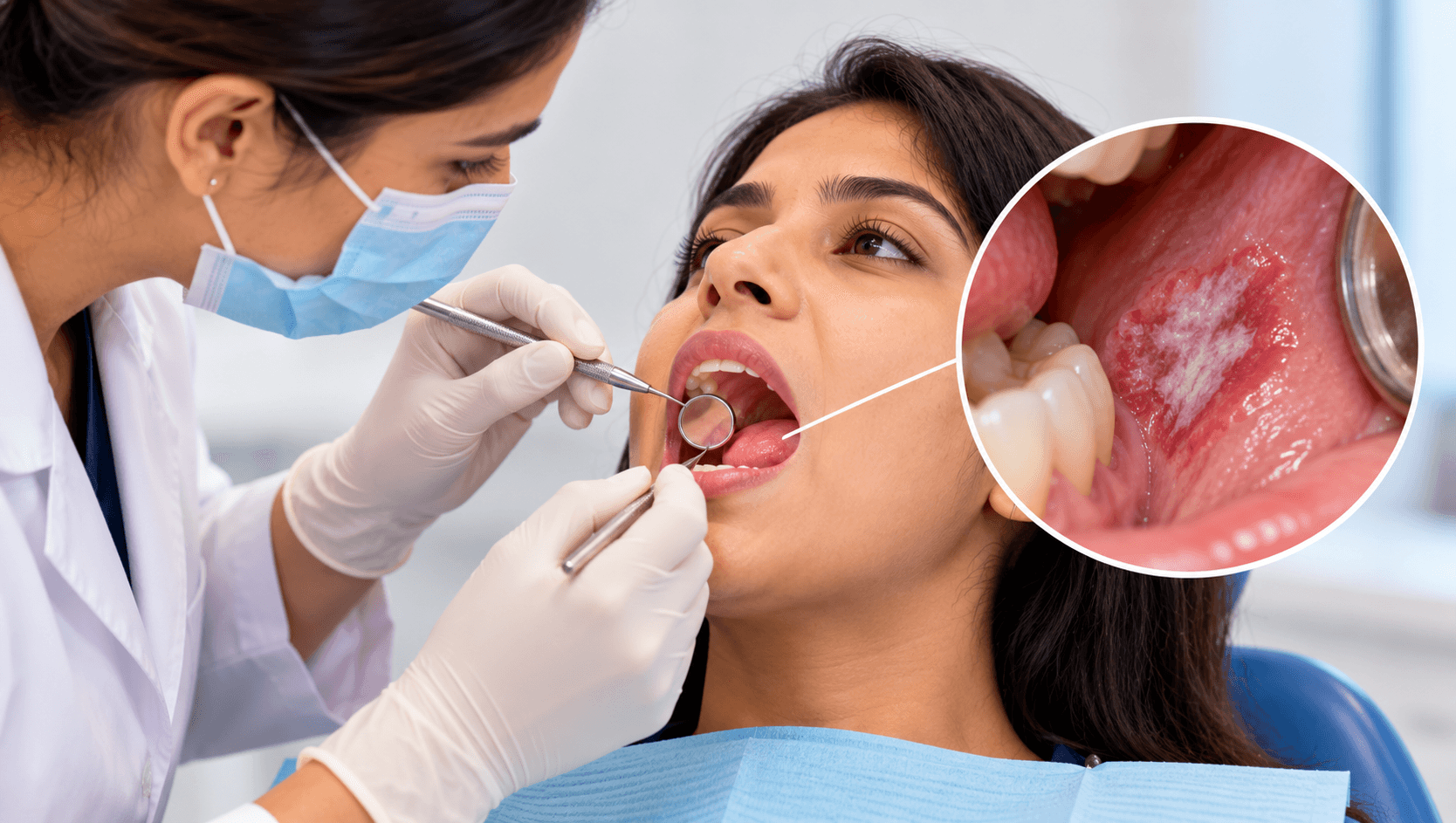
Oral cancer is among the most common cancers in India and across South and Southeast Asia, and it is one of the few malignancies that is directly visible to a clinician performing a routine examination.
Despite that, most cases are diagnosed at an advanced stage, and the five-year survival figures reflect it. The reason is rarely that the lesion was invisible. It is that it was not looked for, or that it was seen and watched for too long.
This guide covers the lesions that carry malignant potential, how to examine for them, when to biopsy rather than review, and how to follow patients up.
What OPMD means
An oral potentially malignant disorder is a clinical presentation that carries a risk of malignant transformation, either at the site of the lesion or elsewhere in clinically normal-appearing mucosa.
The second half of that definition matters. Field cancerisation means the entire mucosa of a patient exposed to tobacco, areca nut and alcohol is at risk, not only the visible lesion. A patient whose leukoplakia is excised remains at elevated risk across the whole oral cavity, which is why follow-up continues after apparently successful treatment.
The main disorders are leukoplakia, erythroplakia, erythroleukoplakia, oral submucous fibrosis, oral lichen planus and lichenoid lesions, proliferative verrucous leukoplakia, actinic cheilitis, palatal lesions in reverse smokers, and oral lesions of discoid lupus erythematosus. Certain inherited conditions, including dyskeratosis congenita and Fanconi anaemia, also carry elevated risk.
The individual disorders
Leukoplakia is a predominantly white plaque of questionable risk, having excluded other known diseases or disorders that carry no increased risk of cancer. It is a diagnosis of exclusion - you must rule out frictional keratosis, candidiasis, lichen planus, leukoedema, white sponge naevus and others first. Homogeneous leukoplakia is thin, uniform and lower risk. Non-homogeneous leukoplakia - speckled, nodular, verrucous or with a red component - carries substantially higher risk.
Erythroplakia is a fiery red patch that cannot be characterised as any other definable disease. It is uncommon and it is the most dangerous of these lesions: a high proportion show severe dysplasia, carcinoma in situ or invasive carcinoma at first biopsy. An erythroplakic lesion is a biopsy, not a review.
Oral submucous fibrosis is strongly associated with areca nut and gutkha use and is highly prevalent in India and South Asia. It presents with burning on spicy food, blanching and marbling of the mucosa, loss of elasticity, palpable fibrous bands, and progressive trismus. It is chronic, progressive and carries significant malignant potential. Trismus in a patient who chews areca nut is a presenting sign, not an incidental finding.
Oral lichen planus is relatively common; the reticular form with Wickham's striae is usually asymptomatic, while erosive, atrophic and ulcerative forms are symptomatic and carry greater risk. Transformation risk overall is low but real, which means these patients need periodic review rather than discharge.
Proliferative verrucous leukoplakia is a distinct and aggressive entity: multifocal, progressive, resistant to treatment, with a high rate of recurrence and transformation. It is often recognised only retrospectively after several years of spreading lesions.
Actinic cheilitis affects the vermilion of the lower lip in patients with chronic sun exposure.
Examining properly
A systematic oral mucosal examination takes about ninety seconds and should be performed on every patient at every recall, not only on those with risk factors.
Follow a fixed sequence so that nothing is omitted: lips and vermilion, labial mucosa, buccal mucosa and vestibules, gingivae, hard and soft palate, oropharynx and tonsillar region, dorsum of tongue, lateral borders of the tongue, ventral tongue, floor of mouth. Finish with bimanual palpation of the floor of mouth and palpation of the cervical lymph nodes.
The lateral border and ventral surface of the tongue and the floor of mouth are the highest-risk sites and the ones most often skipped, because they require you to grasp the tongue with gauze and retract it. Do that on every patient.
Take a proper risk history: tobacco in all forms including smokeless, areca nut, gutkha and pan masala, alcohol, and - relevant for oropharyngeal sites - HPV risk. Record quantity and duration, not merely yes or no.
Record any lesion with site, size, colour, surface, borders, consistency and symptoms, and photograph it. A photograph in the record is worth more than a paragraph of description when you are deciding six weeks later whether something has changed.
When to biopsy
The default should be biopsy, with watchful waiting the exception that requires justification.
Biopsy immediately, or refer for immediate biopsy, if the lesion is erythroplakic or has a red component; is non-homogeneous, speckled, verrucous, nodular or indurated; is ulcerated without an identifiable cause; is at a high-risk site such as the lateral or ventral tongue, floor of mouth or soft palate complex; is associated with lymphadenopathy; or occurs in a patient with a previous oral malignancy.
The widely used rule that any ulcer persisting beyond two weeks without an obvious cause requires referral remains sound.
Where a cause is identifiable and removable - a sharp cusp, an overextended denture flange, a cheek-biting habit - remove the cause and review in two weeks. If the lesion has not resolved, biopsy it. The removable-cause pathway is legitimate; what is not legitimate is repeated reviews of an unchanged lesion over months.
Incisional biopsy should sample the most suspicious area, including the red or indurated component rather than the convenient white edge, and should include a margin of adjacent normal tissue with adequate depth. Multiple site biopsies may be needed for large or multifocal lesions. Adjunctive techniques such as toluidine blue, autofluorescence devices and brush cytology may assist in site selection but do not replace scalpel biopsy and histopathology.
Interpreting the report and managing the patient
Epithelial dysplasia is graded histologically, commonly as mild, moderate or severe, with carcinoma in situ at the extreme. Higher grade is associated with higher transformation risk, but the correlation is imperfect: lesions without dysplasia do transform, and some dysplastic lesions regress. Grading informs management; it does not determine outcome.
Management combines three elements.
Removing the cause. Tobacco and areca nut cessation is the single most effective intervention available, and it is the one most often reduced to a one-line note. It needs active support, referral where available, and repeated discussion. Some lesions regress entirely on cessation.
Treating the lesion. Surgical excision, laser excision or ablation, or cryotherapy, depending on grade, site, size and the patient. Importantly, excision does not eliminate risk - recurrence and transformation at the same or other sites remain possible, which is why follow-up continues.
Follow-up. Lifelong, at intervals determined by lesion type and dysplasia grade, with photographs at each visit for comparison. Patients should also be taught to self-examine and to report changes between appointments.
For oral submucous fibrosis specifically, management includes absolute areca nut cessation, mouth-opening exercises and physiotherapy, and medical or surgical intervention for severe trismus - alongside the recognition that progressive trismus eventually prevents adequate examination of the very mucosa that needs monitoring.
Making this routine
Three habits change outcomes more than any device.
Examine every patient's mucosa systematically, every time, including the tongue borders and floor of mouth.
Ask about tobacco and areca nut use properly, record it, and act on it. In India in particular, the highest-risk patients in your chair are frequently not the ones you would predict from appearance.
Biopsy or refer rather than review when the lesion has any high-risk feature. The cost of an unnecessary biopsy is small; the cost of a delayed diagnosis is measured differently.
Our Oral Pathology Course Online covers oral disease diagnosis and management, and our Online Oral Medicine Course covers oral diagnosis and lesion assessment. Our Oral Pathology and Oral Diagnosis Test Series cover these topics for licensing and postgraduate examinations.
Related reading: our articles on dental records and documentation, and dental treatment planning.