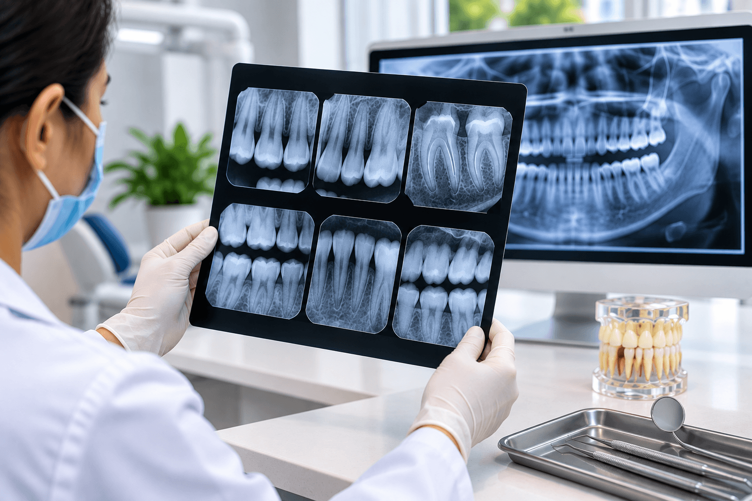
Dental Radiography: Understanding Intraoral X-Rays for Accurate Diagnosis
A clinical examination provides important information about a patient's oral health, but some dental conditions cannot be completely evaluated by visual examination alone. Dental radiography provides dentists with additional diagnostic information by allowing them to examine structures that may not be visible during a routine clinical examination.
Intraoral X-rays are commonly used in dental practice to evaluate teeth, supporting bone, roots, and surrounding structures. When combined with the patient's history and clinical findings, dental radiographs can contribute significantly to diagnosis and treatment planning.
The purpose of dental radiography is not simply to obtain an image. The radiograph needs to be selected, positioned, processed, and interpreted appropriately so that the information obtained is clinically useful.
Clinical Question → Radiographic View → Diagnosis
A useful way to understand dental radiography is to begin with the clinical question rather than simply taking an X-ray without a specific purpose.
Clinical Question
Before prescribing a radiograph, the dentist should consider what information is required.
For example:
Is there suspected dental caries? Is the root structure being evaluated? Is there bone loss? Is an unerupted tooth present? Is there a periapical lesion? Is the patient undergoing endodontic treatment? Is additional information required before restorative or surgical treatment?
Identifying the clinical question helps determine which radiographic examination is appropriate.
Bitewing Radiographs
Bitewing radiographs are particularly useful for evaluating the crowns of posterior teeth and the interproximal areas.
They can assist in detecting:
Interproximal dental caries Recurrent caries around restorations Crestal bone levels Certain changes that may not be clearly visible during clinical examination
Proper positioning is important because overlapping contacts can make interpretation more difficult.
Periapical Radiographs
A periapical radiograph provides a view of the entire tooth, including the crown, root, and surrounding periapical structures.
It may be useful when evaluating:
Root morphology Periapical pathology Endodontic conditions Dental trauma Localized periodontal or bone changes Teeth with suspected infection
Periapical imaging can therefore provide valuable information when the clinical examination suggests that pathology may be present around the root or surrounding bone.
Occlusal Radiographs
Occlusal radiographs provide a broader view of a particular region of the dental arch. Depending on the clinical situation, they may assist in evaluating the position of unerupted teeth, larger areas of the jaw, or certain dental and bony conditions.
Although modern dental imaging has expanded considerably, understanding the indications and limitations of different conventional radiographic views remains an important part of dental education.
Reading a Dental X-Ray: What Should Dentists Look For?
Radiographic interpretation should be systematic rather than limited to the area where a problem is suspected.
A dentist may evaluate:-
Teeth: Look at tooth structure, restorations, caries, root morphology, and other visible abnormalities.
Periodontal Structures: Assess relevant bone levels and supporting structures.
Periapical Region: Examine the area around the roots for radiographic changes.
Developing or Unerupted Teeth: Consider tooth position, eruption pattern, and relationship with surrounding structures.
Adjacent Structures: Depending on the radiograph, surrounding anatomical structures may also need to be assessed.
A systematic approach reduces the possibility of overlooking clinically relevant findings.
Dental Radiography in Treatment Planning
Radiographs can play an important role in dental treatment planning. Before beginning certain procedures, dentists may require additional information about the tooth and surrounding structures.
For example, radiographic information may contribute to planning for:-
Root canal treatment Dental extractions Restorative procedures Periodontal treatment Assessment of impacted teeth Evaluation of dental trauma Certain surgical procedures
Radiographs should complement—not replace—clinical examination. The final diagnosis should be based on the overall clinical picture.
A Simple Radiographic Diagnosis Workflow
1. Identify the Clinical Problem Understand the patient's symptoms and clinical findings.
2. Select the Appropriate Radiograph Choose the imaging technique that can provide the required diagnostic information.
3. Obtain a Quality Image Use appropriate positioning and exposure techniques.
4. Examine the Entire Radiograph Evaluate the relevant teeth, supporting structures, and surrounding anatomy.
5. Correlate With Clinical Findings Compare the radiographic appearance with the patient's history and clinical examination.
6. Use the Findings for Treatment Planning Incorporate relevant radiographic information into the overall diagnosis and treatment plan.
Why Dental Radiology Knowledge Matters
Understanding dental radiology and diagnostic imaging is an important clinical skill for dentists. Correct interpretation of radiographs requires knowledge of normal anatomy as well as recognition of abnormal radiographic appearances.
For dental graduates, developing radiographic interpretation skills can improve confidence when assessing routine and complex cases. Practical exposure, case discussions, and continued dental education can further strengthen these diagnostic abilities.
At Master Clinical Dentistry Institute (MCDI), dental education focuses on developing clinical knowledge and practical decision-making skills that can be applied in everyday dental practice.
Key Takeaways
Dental radiography provides diagnostic information that may not be available through clinical examination alone.
The choice of radiograph should be based on the specific clinical question.
Bitewing radiographs are useful for evaluating posterior crowns, interproximal areas, and crestal bone levels.
Periapical radiographs provide information about the entire tooth, roots, and surrounding periapical structures.
Radiographic interpretation should be systematic and include the entire relevant image.
Image quality and correct positioning are important for obtaining useful diagnostic information.
Dental X-rays can contribute significantly to diagnosis and treatment planning.
Radiographic findings should always be interpreted alongside the patient's history and clinical examination.
Frequently Asked Questions
What is dental radiography? Dental radiography is the use of X-ray imaging to obtain information about teeth, bone, roots, and other oral structures that may not be fully visible during clinical examination.
What is an intraoral X-ray? An intraoral X-ray is a dental radiograph in which the image receptor is positioned inside the patient's mouth to capture detailed images of teeth and surrounding structures.
What are bitewing X-rays used for? Bitewing radiographs are commonly used to examine the crowns of posterior teeth, detect interproximal caries, evaluate restorations, and assess crestal bone levels.
What is a periapical X-ray used for? A periapical radiograph captures the complete tooth and surrounding periapical region and can be useful for evaluating root and periapical conditions.
Why is radiographic interpretation important for dentists? Correct interpretation helps dentists correlate imaging findings with clinical information and make better-informed diagnostic and treatment-planning decisions.
Conclusion
Dental radiography is an important component of modern dental diagnosis. Intraoral X-rays can provide valuable information about tooth structure, roots, supporting bone, and surrounding tissues that may not be visible during a routine examination.
The effectiveness of dental X-ray diagnosis depends on appropriate image selection, correct positioning, good image quality, and systematic interpretation. Most importantly, radiographic findings should always be considered together with the patient's history and clinical examination.
Developing strong dental radiology and diagnostic skills can help dentists approach patient assessment and treatment planning more effectively. Continued education and practical clinical training can further strengthen these essential skills.