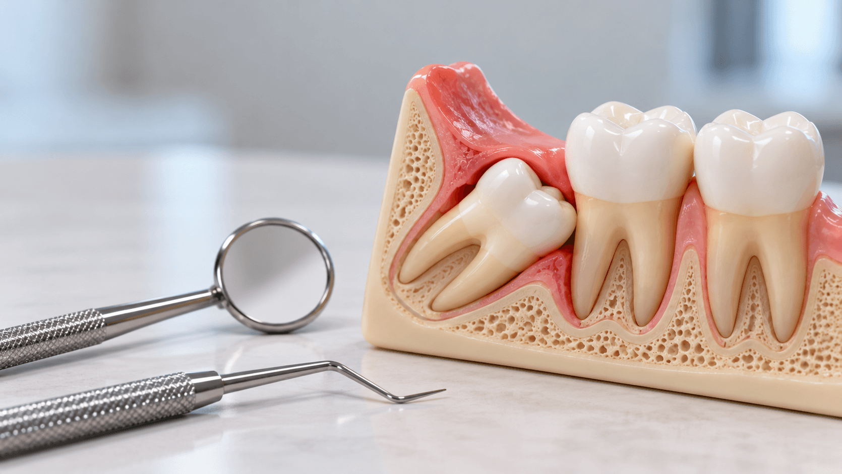
Third molar surgery is the commonest minor oral surgical procedure in general dental practice and the one that most often goes from routine to difficult without warning.
The difference between a twenty-minute extraction and a ninety-minute struggle is almost entirely decided before the scalpel is picked up - in the radiographic assessment, in the honest estimate of difficulty, and in the decision about whether this particular tooth belongs in your chair at all.
This guide covers assessment, technique and the complications you must be able to manage.
Indications and the case for leaving it alone
Not every impacted third molar needs removing, and routine prophylactic removal of asymptomatic, disease-free impacted third molars is not supported by current guidance.
Accepted indications include recurrent pericoronitis, caries in the third molar or distal caries in the second molar attributable to it, periodontal pocketing distal to the second molar, resorption of the second molar root, associated cyst or tumour, fracture of the mandible through the tooth, as part of orthodontic or orthognathic planning, and a tooth beneath a planned prosthesis.
The decision is clinical, and the conversation with a patient who has an asymptomatic impaction should be about surveillance rather than automatic removal.
Age matters. Removal is easier and complications are fewer in younger patients, where roots are incompletely formed and bone is more elastic. A patient in their forties with a deeply impacted tooth and fully formed roots close to the canal is a different proposition from a nineteen-year-old with the same radiographic picture.
Assessment and classification
Two classifications remain in routine use and are examined everywhere.
Winter's classification describes the angulation of the impacted tooth relative to the long axis of the second molar: mesioangular, distoangular, horizontal, vertical, buccoangular, linguoangular and inverted.
The practical ranking of difficulty is counterintuitive and worth remembering: mesioangular impactions are generally the least difficult, and distoangular the most, because the path of withdrawal of a distoangular tooth runs into the ascending ramus.
Pell and Gregory classification describes the relationship to the ramus and the depth relative to the occlusal plane. Class 1, 2 and 3 describe available space between the second molar and the ramus; Position A, B and C describe depth, with C being deepest.
Beyond classification, assess root morphology and number, degree of root formation, bone density, the periodontal ligament space, the relationship to the second molar, the proximity to the maxillary sinus for upper teeth, and - most importantly for lower teeth - the relationship to the inferior alveolar canal.
Seven radiographic signs on a panoramic film suggest a close relationship with the canal: darkening of the root where it crosses the canal, deflection of the root, narrowing of the root, a dark and bifid root apex, interruption of the white line or lines of the canal, diversion of the canal, and narrowing of the canal. Interruption of the cortical white line, diversion and narrowing of the canal are the signs most strongly associated with true proximity.
When one or more of these signs is present, CBCT is justified - not to change whether you operate, but to determine the three-dimensional relationship, which influences technique and consent. It is not justified routinely for every third molar.
Technique
Anaesthesia: inferior alveolar and lingual nerve block with long buccal infiltration for lower teeth; posterior superior alveolar and greater palatine or infiltration for upper.
Flap design: a buccal envelope flap extending from the distal of the second molar with a distobuccal relieving incision is standard. The distal incision must angle buccally, away from the lingual plate, to protect the lingual nerve. A three-cornered flap with a mesial relieving incision gives wider access for deeper impactions.
Principles that apply to every flap: broad base for blood supply, full thickness raised cleanly from bone, margins resting on sound bone at closure, and no tension.
Bone removal: a surgical handpiece with copious irrigation. Never use an air-driven handpiece that exhausts air into the surgical field, because of the risk of surgical emphysema. Remove buccal and distal bone to expose the crown to its maximum convexity and create a point of application.
Sectioning: often faster and less traumatic than removing more bone. Divide crown from roots, or section the crown, or divide fused roots, according to the impaction. The general rule is that it is better to section the tooth than to remove the bone around it.
Elevation: use controlled, deliberate force with a defined fulcrum and a finger supporting the mandible. The adjacent second molar is not a fulcrum.
Debridement: remove the follicle, irrigate thoroughly, check for loose bone fragments, smooth sharp margins.
Closure: sutures as required for flap position and haemostasis. Our Dental Suturing Course covers the technique specifically.
Coronectomy
Where the radiographic and CBCT findings indicate a genuinely intimate relationship between the roots and the inferior alveolar canal, coronectomy - deliberate removal of the crown with retention of the roots - is an accepted alternative that substantially reduces the risk of nerve injury.
Conditions for it: the tooth must be vital and non-infected, the roots must not be mobilised during the procedure, and the patient must accept the need for long-term review and the possibility of later root migration requiring removal.
It is not a salvage technique for a procedure that has gone wrong, and it should be planned and consented in advance rather than decided mid-operation.
Complications
Alveolar osteitis, or dry socket, typically presenting two to four days postoperatively with severe pain and a denuded socket. Risk factors include smoking, oral contraceptive use, surgical trauma and poor oral hygiene. Management is irrigation, a sedative dressing and analgesia; it is not an antibiotic indication in the absence of spreading infection.
Inferior alveolar nerve injury, giving altered sensation of the lower lip and chin. Most are temporary; a minority are permanent. Risk is predicted by the radiographic signs above and by surgical technique.
Lingual nerve injury, giving altered sensation and taste on the tongue. Avoided by keeping instruments away from the lingual plate, by correct distal incision angulation, and by careful elevation.
Trismus, swelling and pain, which are expected rather than complications in the strict sense, but which should be proportionate and improving by day three.
Infection, bleeding, and in upper teeth oroantral communication and displacement of a root into the sinus.
Fracture of the mandible, rare, more likely in deeply impacted teeth in older patients with atrophic mandibles.
Displacement of a lower third molar into the submandibular space.
Damage to the adjacent second molar, which is more common than reported and is usually the result of using it as a fulcrum.
Document the nerve injury risk discussion in your notes before every lower third molar removal, with the specific radiographic findings you based it on. This is the single most frequent subject of complaint and litigation in minor oral surgery.
Knowing when to refer
There is no credit for struggling. Refer when the radiographic signs indicate genuine canal proximity and you are not trained in coronectomy; when the impaction is deep, distoangular and Class 3 Position C; when the patient has a medical history that complicates surgery, such as antiresorptive medication, anticoagulation or immunosuppression; when previous attempts have failed; and when the time required would exceed what you can safely manage in your setting.
The skill to develop first is accurate difficulty assessment. A clinician who correctly predicts which cases they should not attempt will have a better complication record than one who is technically faster but takes on everything.
Our Oral Surgery Course for Dentists covers minor oral surgery and extraction hands-on, and our Online Oral Surgery Course covers exodontia, impactions and surgical planning remotely. Our Dental Radiology and CBCT courses cover the radiographic assessment this procedure depends on.
Related reading: our articles on local anaesthesia techniques, suturing in dentistry, and medical emergencies in dental practice.