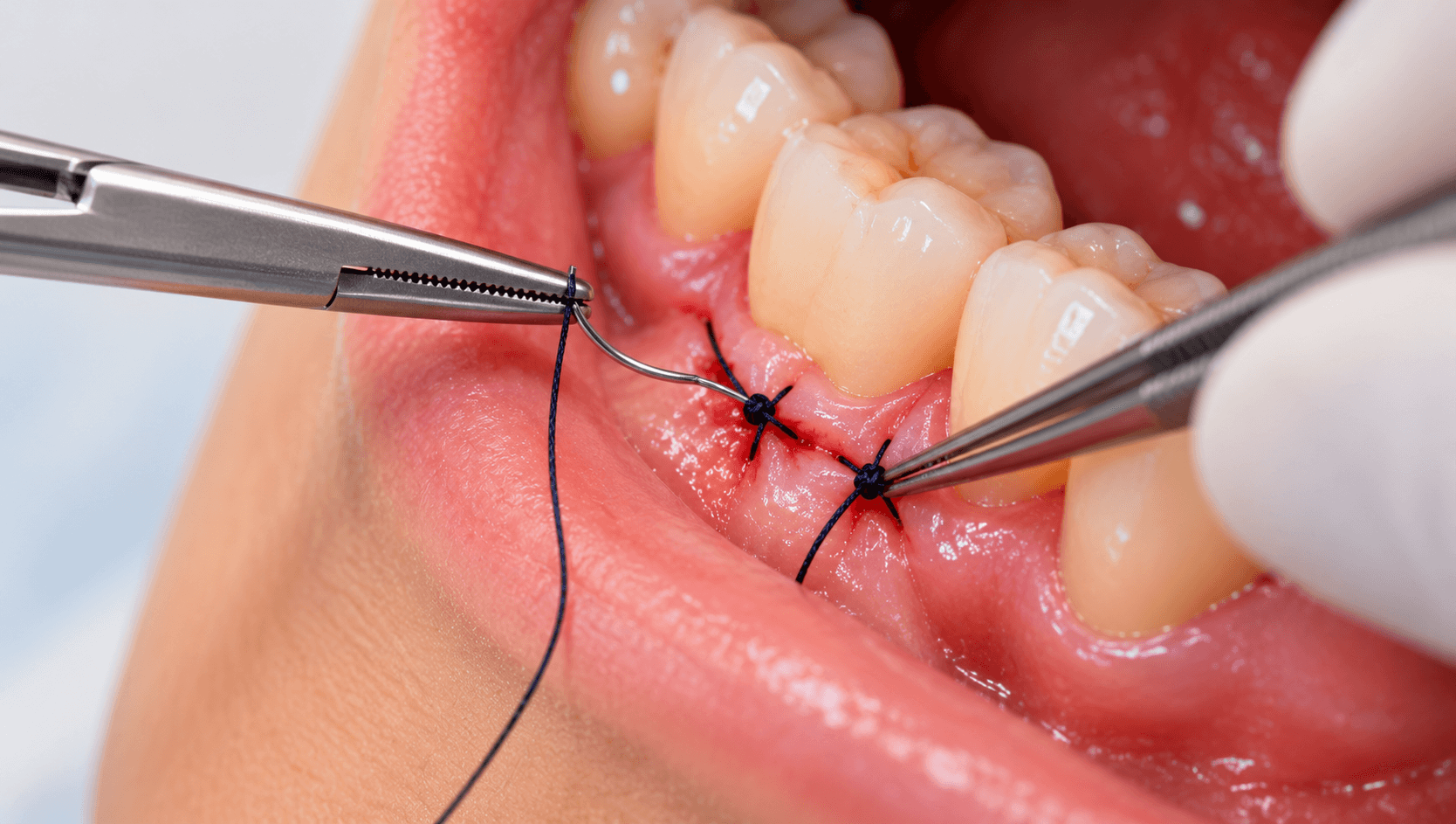
Suturing is taught quickly, practised rarely during undergraduate training, and then performed under pressure at the end of a long surgical procedure when concentration is lowest.
It is also the step that determines how the wound heals. A technically excellent extraction or flap procedure closed badly produces dehiscence, delayed healing, pain and sometimes graft loss. The converse is rarely true - good closure cannot rescue bad surgery - but poor closure routinely spoils good surgery.
This guide covers material selection, needles, the techniques that matter in dentistry, and the handling details that separate a secure closure from one that comes apart on day two.
What sutures are actually for
Four functions, and it helps to be explicit about which one you need in a given case.
Approximating the wound edges in the correct position, passively and without tension.
Maintaining a blood clot and achieving haemostasis.
Stabilising flap position, particularly where the flap has been repositioned coronally, apically or laterally.
Protecting a graft or membrane beneath.
The most important principle follows from the first: sutures approximate, they do not close under tension. A flap pulled together by its sutures will blanch, the tissue will necrose at the margin, and the wound will open. If the flap does not sit passively in position before you suture, release it properly with a periosteal releasing incision rather than relying on thread.
Choosing a material
Resorbable or non-resorbable is the first decision.
Resorbable materials - polyglactin 910, polyglycolic acid, catgut and chromic catgut - do not require removal, which suits paediatric patients, anxious patients, inaccessible sites and patients unlikely to return. Resorption times differ substantially between materials; catgut degrades quickly and unpredictably, synthetic polymers more slowly and more reliably.
Non-resorbable materials - silk, nylon, polypropylene, PTFE - require removal but offer predictable tensile strength for as long as they are in place.
Monofilament or multifilament is the second decision, and it matters more than most clinicians realise.
Braided multifilament materials such as silk and polyglactin handle beautifully and hold knots securely, but their interstices wick fluid and harbour bacteria. Plaque accumulates on them rapidly in the mouth.
Monofilament materials such as nylon, polypropylene and PTFE do not wick, accumulate far less plaque and cause less tissue reaction, but they have memory, handle stiffly and require more throws to hold a knot securely.
In practice: silk remains common for routine extraction sockets because it handles well and is inexpensive, and it is removed within a week. PTFE is the preferred material around implants, grafts and membranes, where plaque accumulation matters most and the suture may stay longer. Polyglactin is the standard resorbable choice. Choose the material for the site and duration, not out of habit.
Size in dentistry is usually 3-0 to 5-0, with the convention that higher numbers mean finer thread. A 3-0 for routine extraction sockets, 4-0 for most flap work, 5-0 for anterior aesthetic sites and delicate papillae.
Needles
Reverse cutting needles are used for most oral work. The cutting edge is on the outer convexity of the curve, which reduces the risk of the suture tearing through toward the wound edge. Conventional cutting needles, with the edge on the inner curve, are more likely to cut out.
Taper-point needles are for delicate, easily penetrated tissue and are less commonly indicated in oral mucosa and gingiva.
Curvature is typically three-eighths or one-half circle. The half-circle needle is easier to use in the posterior mouth where access is limited.
Grasp the needle with the needle holder approximately one-third to one-half of the way back from the tip - not at the swage, which weakens it, and not at the tip, which bends it. Rotate the needle through the tissue along its curve rather than pushing it straight; forcing a curved needle in a straight line tears tissue and bends needles.
The techniques that matter
Simple interrupted. The default. Each suture is independent, so failure of one does not compromise the rest, and tension can be adjusted at each site. Used for extraction sockets, relieving incisions and most flap margins.
Figure-of-eight. A single suture crossing over the socket, useful for retaining a dressing or haemostatic material in an extraction socket.
Sling or suspensory suture. Passed around the neck of a tooth to suspend a flap, used where one flap needs positioning independently of the tissue on the opposite side - common in periodontal surgery and in single-sided flaps.
Horizontal mattress. Everts the wound edges, distributes tension across a broader area, and improves haemostasis. Useful for flap closure over grafts.
Vertical mattress. Provides both deep and superficial approximation with good eversion, and is valuable where precise edge adaptation matters.
Continuous suture. Faster for long incisions and distributes tension evenly, but a single failure can compromise the whole line. Continuous locking variants add security.
Papilla-based and modified techniques in periodontal plastic surgery, where preservation of the interdental papilla is the objective.
Two rules apply to all of them. Pass the needle from the thinner, more mobile tissue to the thicker, less mobile tissue - usually buccal to lingual, and from free flap to attached tissue. And take an adequate bite, roughly 2 to 3 mm from the wound edge, into firm tissue; a bite taken too close to the margin cuts through.
Knots and tension
The surgeon's knot - a double throw first, then a single throw in the opposite direction, then a further single throw - is the standard in oral surgery. The double first throw provides friction that holds approximation while you lay the second throw.
Alternate the direction of each throw. Throws laid in the same direction produce a slip knot that loosens.
Lay the knot flat rather than pulling it into a bunch, and position it to the side of the incision line, not over it. A knot sitting on the wound margin acts as a plaque trap directly where healing is happening.
On tension: the tissue should be approximated, not blanched. If the tissue blanches as you tighten, the suture is too tight and the margin will necrose. This is the single commonest error in dental suturing, and it is nearly always a consequence of trying to close a flap that was never released adequately.
Monofilament materials need more throws than braided ones to hold securely - plan for five or six on PTFE and nylon.
Aftercare and removal
Give clear post-operative instructions: no rinsing for the first day, then gentle warm saline or chlorhexidine rinses, no smoking, soft diet, and avoidance of disturbing the area with the tongue or a toothbrush while still cleaning the rest of the mouth.
Non-resorbable sutures in the mouth are generally removed around five to seven days, earlier in the anterior aesthetic zone and sometimes later where a graft needs prolonged stabilisation. Oral mucosa heals quickly, and sutures left beyond ten days accumulate plaque and cause inflammation of their own.
To remove: clean the area, grasp the knot, cut the suture below the knot on the side closest to the tissue, and draw it out so that no portion that was exposed to the oral environment passes through the tissue.
Count them. Record the number placed and the number removed. A retained suture is a common and entirely avoidable cause of a persistent inflammatory lesion that gets investigated as something more serious.
Our Dental Suturing Course offers a hands-on suture workshop, and the Online Dental Suturing Course covers materials, needles and technique remotely. Both pair naturally with our Oral Surgery and Periodontology courses.
Related reading: our articles on impacted third molar surgery and sinus lift and bone grafting, where closure technique determines the outcome.