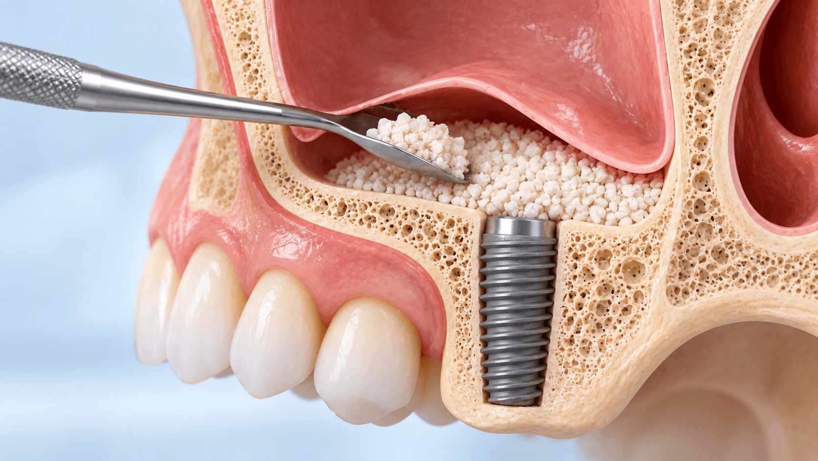
The posterior maxilla is where implant planning most often stops. Sinus pneumatisation and alveolar resorption leave residual bone that will not accept a fixture, and the clinician's options narrow to augmenting the site, going around it, or declining the case.
Grafting is the most commonly chosen answer, and it is a well-evidenced one. It is also a procedure where the gap between the textbook description and the clinical reality is wide, mostly because of what happens when the membrane tears.
This guide covers the decision thresholds, the two main sinus techniques, the graft materials and the complications - and is explicit about when not to graft.
The decision: how much bone, and where
The governing measurement in the posterior maxilla is residual bone height below the sinus floor, measured on CBCT, not on a panoramic radiograph.
As a working framework, used alongside bone quality and width rather than instead of them: with generous residual height, conventional implant placement without augmentation is possible. With moderate residual height, a crestal or transalveolar approach is usually feasible, often with simultaneous implant placement, because there is enough bone for primary stability. With minimal residual height, a lateral window approach is generally indicated, and the choice between simultaneous and staged placement turns on whether primary stability can be achieved.
Width matters independently. Adequate height with a knife-edge ridge is a horizontal augmentation problem, not a sinus problem, and the two are frequently confused in treatment plans.
Assess the sinus itself before planning anything: septa, mucosal thickening, patency of the ostium, and any history of chronic sinusitis. A patient with an obstructed ostium should not have a graft placed into that sinus until the obstruction is addressed.
The lateral window technique
A mucoperiosteal flap is raised, a window prepared in the lateral maxillary wall - with a round bur under copious irrigation, or with piezosurgery, which is considerably more membrane-friendly - and the Schneiderian membrane carefully elevated from the sinus floor and walls. The elevated space is filled with graft material, the window may be covered with a resorbable membrane, and the flap closed without tension.
The technical points that determine success: design the window to give access to the whole floor without extending unnecessarily; identify septa on CBCT beforehand, because a septum encountered unexpectedly is the commonest cause of perforation; elevate slowly and keep instruments in contact with bone, not membrane; and release the membrane fully at the mesial and distal extent before placing graft, or it will tear under the pressure of the material.
Simultaneous implant placement is appropriate where residual bone gives primary stability. Where it does not, stage the case, typically allowing several months of graft maturation before placement. Forcing simultaneous placement into inadequate bone produces a mobile implant in a graft, which is a failure with a long recovery.
The crestal or transalveolar approach
Where residual height is moderate and the lift required is modest, the sinus floor can be elevated through the implant osteotomy itself.
The original osteotome technique uses increasingly wide osteotomes to greenstick-fracture and elevate the floor, with graft introduced through the osteotomy. Variations using hydraulic pressure, specialised drills with reverse cutting geometry, or balloon devices reduce the percussive element, which patients appreciate and which lowers the risk of benign paroxysmal positional vertigo.
The advantages are a smaller flap, less morbidity, less swelling and a shorter procedure. The limitation is that you are working blind: you cannot see the membrane, so perforation is detected indirectly, usually with the Valsalva manoeuvre, and the achievable lift is smaller.
It is the right choice for the modest lift in adequate bone, and the wrong choice for the severely pneumatised sinus with minimal residual height, however appealing the lower morbidity sounds.
Graft materials
Autogenous bone is osteogenic, osteoinductive and osteoconductive, and remains the biological reference standard. Its disadvantages are donor site morbidity, limited volume, and a resorption rate that can be substantial.
Allograft, from human donors, processed as freeze-dried or demineralised freeze-dried bone. Osteoconductive and to a degree osteoinductive, with no donor site.
Xenograft, most commonly deproteinised bovine bone mineral. Osteoconductive with very slow resorption, which gives excellent volume maintenance - the property that makes it the workhorse material for sinus grafting. The trade is that residual particles persist long term.
Alloplast: synthetic materials such as beta-tricalcium phosphate, hydroxyapatite and bioactive glass. Osteoconductive, unlimited supply, no biological donor concerns.
In practice composite grafts are common, combining a xenograft for volume stability with autogenous or allograft for biological activity. For the sinus specifically, the enclosed five-walled defect is highly favourable and good results are achieved with a wide range of materials, which is why material choice matters less here than in horizontal ridge augmentation.
Horizontal and vertical ridge augmentation
Grafting the sinus is the easy augmentation. Rebuilding a ridge is harder, because the defect is not enclosed.
Guided bone regeneration uses a particulate graft stabilised beneath a barrier membrane, resorbable or non-resorbable, to exclude soft tissue while bone forms. Success depends on space maintenance, graft stability, primary wound closure without tension, and an intact membrane.
Block grafts, autogenous or allogeneic, provide structural volume for larger horizontal defects and are rigidly fixed with screws.
Ridge splitting is an option where width is deficient but there is sufficient height and a cancellous core between the plates.
Vertical augmentation is the most technically demanding and has the highest complication rate. Tension-free primary closure over a vertically augmented site is difficult, and wound dehiscence with graft exposure is the dominant failure mode.
The general rule holds throughout: horizontal augmentation is predictable, vertical augmentation is not, and the difficulty is soft tissue management rather than the graft itself.
Complications and how to handle them
Schneiderian membrane perforation is the most common intraoperative complication. Small perforations can often be managed by continuing to elevate carefully until the tear is surrounded by elevated membrane, then covering it with a resorbable collagen membrane before grafting. Large perforations that cannot be reliably covered are an indication to abandon the graft, close, and return after healing. A graft placed under a perforated membrane that was not properly managed will become infected.
Postoperative sinusitis, presenting with pain, congestion, purulent discharge and sometimes fever. More likely with pre-existing sinus disease, ostium obstruction, perforation or graft contamination.
Graft infection, which usually requires removal of the graft.
Wound dehiscence and graft exposure, particularly after ridge augmentation. Exposed non-resorbable membranes become colonised and generally need removal.
Bleeding from a branch of the posterior superior alveolar artery running within the lateral wall - identifiable on CBCT and avoidable with window design.
Benign paroxysmal positional vertigo after osteotome malleting.
Two things reduce all of these: CBCT planning that is actually examined rather than glanced at, and operating slowly.
When not to graft
Active sinus pathology or an obstructed ostium should be treated, with ENT involvement, before any graft.
Heavy smokers have materially worse outcomes in all augmentation procedures, and vertical augmentation in a heavy smoker is a poor use of everyone's time.
Uncontrolled systemic disease, particularly poorly controlled diabetes.
Patients on antiresorptive or antiangiogenic medication require careful assessment for medication-related osteonecrosis risk.
And there is the strategic question: is a graft the right answer at all? Short implants in the available bone, tilted implants avoiding the sinus, pterygoid implants, zygomatic implants in the severely atrophic maxilla, or a well-made removable prosthesis are all legitimate alternatives. A patient who wants to avoid nine months of staged grafting is not being unreasonable, and the graftless options exist precisely for them.
Our Advanced Dental Implant Course covers bone grafting, sinus lift and guided surgery hands-on; the online version covers the same planning content remotely. For the graftless alternatives see our Zygomatic Implant and Basal Implant courses.
Related reading: our guide to zygomatic implants and the atrophic maxilla, our comparison of basal and conventional implants, and our complete guide to modern implant dentistry.