A patient reports clicking, intermittent jaw pain and headaches. Previous advice has ranged from do nothing to it is your bite, with an occlusal adjustment somewhere in between.
Temporomandibular disorders are common, usually self-limiting, and almost always best managed conservatively. They are also the area of dentistry where irreversible treatment is most often performed on inadequate evidence - occlusal adjustment, full-mouth rehabilitation and orthodontics undertaken to treat symptoms that would have improved with reassurance and a splint.
This guide covers assessment, conservative management, where splints genuinely help, and the findings that mean this is not a routine TMD at all.
What TMD covers
Temporomandibular disorders are a group of conditions, not a single diagnosis, and the first useful distinction is between muscular and joint problems.
Masticatory muscle disorders: myalgia, local myalgia, myofascial pain with or without referral. These are the most common presentations and generally the most responsive to conservative care.
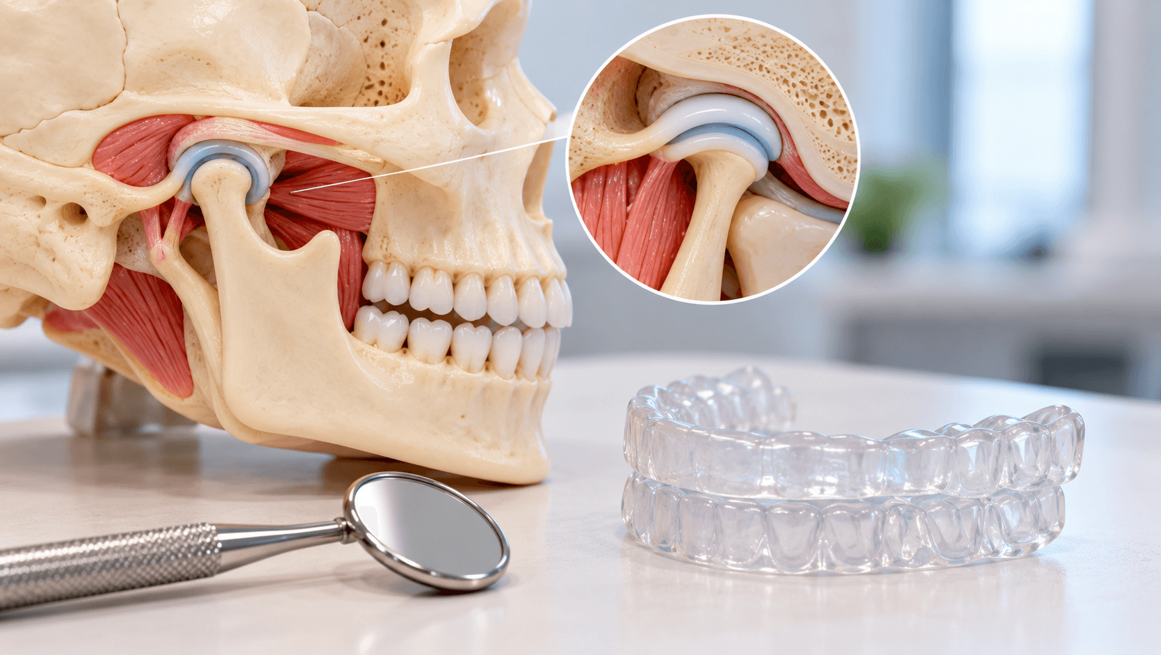
Temporomandibular joint disorders: disc displacement with reduction, with or without intermittent locking; disc displacement without reduction, with or without limited opening; degenerative joint disease; subluxation and dislocation.
Headache attributed to TMD.
A patient frequently has more than one of these, and a muscular component accompanies most joint disorders.
Contributing factors include parafunction, particularly sleep and awake bruxism, trauma, stress and poor sleep, generalised joint hypermobility, and comorbid chronic pain conditions. Occlusion is a far less important factor than a previous generation of teaching suggested, and this is the single most important conceptual correction for clinicians trained on older material.
Taking the history
Characterise the pain: location, onset, duration, quality, severity, what makes it better and worse, diurnal pattern. Pain worse on waking suggests sleep bruxism; pain that builds through the day suggests awake parafunction.
Ask about joint noises - clicking, crepitus - and whether they are associated with pain or with locking. A painless click in a patient with normal function usually requires reassurance and nothing more.
Ask about locking, both closed and open, and whether it resolves spontaneously.
Ask about headache, neck pain, earache, tinnitus and dizziness. Many TMD patients have seen an ENT surgeon first.
Ask about trauma, recent dental treatment including prolonged mouth opening, and general anaesthetic intubation.
Screen for the factors that drive chronic pain: sleep quality, stress, mood, other chronic pain conditions, and medication.
Establish what the patient believes is causing it and what they fear. A substantial part of TMD management is addressing the belief that the joint is degenerating or that surgery is inevitable.
The examination
Measure maximum comfortable opening, maximum assisted opening, and lateral and protrusive excursions. Record the numbers, because change over time is more informative than any single reading.
Observe the opening path for deviation, which is a deflection that corrects, or deflection, which does not.
Palpate the masticatory muscles - masseter, temporalis, and the accessible portions of the pterygoids - and the cervical muscles, recording tenderness and whether palpation reproduces the patient's familiar pain. Reproduction of familiar pain is the diagnostic point, not tenderness alone.
Palpate and auscultate the joints laterally and through the external auditory meatus during opening, closing and excursions, noting click timing and crepitus.
Examine the dentition for wear facets, tooth mobility, buccal mucosal ridging and tongue scalloping - signs of parafunction.
Screen for sleep disordered breathing, because bruxism and obstructive sleep apnoea frequently coexist. See our article on sleep apnoea and the dentist's role.
Imaging is not routinely required. Where it is indicated - suspected degenerative change, trauma, or failure to respond to conservative management - CBCT assesses bony structures and MRI assesses the disc and soft tissues. A panoramic radiograph is a screening view, not a TMJ assessment.
Conservative management
This is first-line for almost every patient, and in the majority it is sufficient.
Explanation and reassurance. Explaining that TMD is common, usually self-limiting, and not a sign of joint destruction has a genuine therapeutic effect. Patients who understand their condition improve more than those who do not.
Self-management: soft diet during flare-ups, avoiding wide opening, chewing gum, nail biting and other parafunction, applying heat or cold, and awareness training for daytime clenching.
Jaw exercises and physiotherapy, including gentle range-of-motion and stretching exercises. Physiotherapy referral is valuable and underused.
Analgesia with non-steroidal anti-inflammatory drugs for a limited period during flare-ups.
Addressing sleep, stress and mood, with referral for cognitive behavioural approaches where chronic pain features are present.
Review at six to eight weeks. Most patients improve substantially within this period on conservative measures alone.
Occlusal splints: what they do and do not do
A full-coverage hard acrylic stabilisation splint, worn at night, is the appropriate appliance for most TMD and bruxism presentations. It is reversible, it protects the dentition from parafunctional wear, and it reduces muscular symptoms in a substantial proportion of patients.
Three design principles matter: full occlusal coverage, so that no teeth over-erupt and no single tooth is loaded; even bilateral contacts in the intercuspal position; and canine or anterior guidance in excursions, with posterior disclusion.
Partial coverage appliances - anterior-only devices and the like - carry a risk of unwanted tooth movement and posterior open bite with unsupervised use, and should not be dispensed casually for long-term wear.
What a splint does not do is permanently reposition the disc, cure the condition, or justify subsequent irreversible occlusal change. A splint that relieves symptoms is evidence that the symptoms are responsive to conservative management, not evidence that the patient needs their occlusion rebuilt.
Which leads to the central caution of this article: do not perform occlusal adjustment, full-mouth rehabilitation or orthodontics to treat TMD. The evidence does not support irreversible occlusal therapy as a treatment for temporomandibular disorders, and a patient whose symptoms persist after an irreversible intervention has both the original problem and a new one.
Red flags and referral
Refer, and in some cases refer urgently, when you find any of the following.
Progressive limitation of opening, or acute closed lock that does not resolve.
Unilateral swelling over the joint, fever, or signs of infection.
Facial or lip numbness, or any other cranial nerve sign.
Weight loss, night pain, or a progressive history suggesting neoplasia - metastatic disease and nasopharyngeal tumours can present as jaw pain.
Trismus of unclear cause, especially with a history of tobacco or areca nut use, where oral submucous fibrosis and malignancy must be excluded.
A joint that has recently changed shape, an anterior open bite developing without explanation, or an occlusion that has shifted - which may indicate condylar resorption or a degenerative or inflammatory arthropathy.
Pain unresponsive to appropriate conservative management after an adequate trial.
Suspected neuropathic facial pain or trigeminal neuralgia, which is a different condition with a different management pathway.
Our Online TMJ Course for Dentists covers TMD diagnosis, orofacial pain and splint therapy, and our Dental Occlusion courses cover occlusal analysis and bite management. Our Obstructive Sleep Apnea courses cover the airway assessment that frequently belongs in these patients.
Related reading: our articles on dental occlusion in diagnosis and treatment planning, oral potentially malignant disorders, and sleep apnoea and the dentist's role.