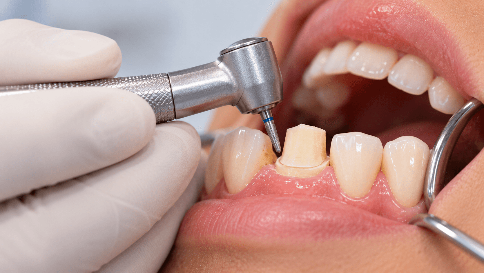
A crown preparation is a series of decisions made with a bur, and most of them are irreversible. The laboratory can compensate for very little, the patient has no way of judging what was done, and the consequences - an unretentive crown, a fractured ceramic, chronic gingival inflammation - usually appear months or years later.
The principles have not changed in decades. What has changed is the materials, and with them the reduction requirements and finish line preferences. A preparation designed for a metal-ceramic crown is not optimal for monolithic zirconia, and a preparation designed for either is wrong for a lithium disilicate onlay.
This guide covers the principles, the material-specific requirements, and the errors that most often cause remakes.
The five objectives
Every decision in a preparation serves one of five objectives, and conflicts between them are what make preparation a matter of judgement.
Preservation of tooth structure. Every micrometre removed is permanent, and pulpal and structural consequences follow from over-reduction.
Retention and resistance form. Retention resists removal along the path of insertion; resistance resists dislodgement by oblique and lateral forces. Resistance is the one that actually fails in practice, and it is governed by preparation height and taper.
Structural durability of the restoration. Adequate reduction so the material has sufficient thickness to resist fracture.
Marginal integrity. A finish line the laboratory can read, the clinician can access, and the material can be finished to.
Preservation of the periodontium. Margin placement, contour and cleansability.
Reduction by material
Reduction requirements are driven by what the restoration is made of, and under-reduction is the commonest cause of a remake.
Metal-ceramic: approximately 1.5 to 2.0 mm occlusal reduction on functional cusps, 1.2 to 1.5 mm axially where porcelain is present, and a shoulder or deep chamfer of around 1.2 to 1.5 mm on the facial. Where metal only, reduction can be considerably less.
Full-contour monolithic zirconia: this is where many clinicians over-prepare out of habit. Monolithic zirconia requires substantially less than metal-ceramic - roughly 1.0 to 1.5 mm occlusally and 0.8 to 1.0 mm axially, with a chamfer finish line. Preparing a monolithic zirconia crown to metal-ceramic dimensions removes sound tooth structure for nothing.
Layered zirconia: more reduction than monolithic, because the veneering porcelain needs space and unsupported porcelain is what chips.
Lithium disilicate full crowns: approximately 1.5 to 2.0 mm occlusally and 1.0 to 1.5 mm axially, with a shoulder or deep chamfer. Lithium disilicate is bonded, so adhesion contributes to retention and the preparation can be more conservative where enamel remains.
Gold: the most conservative of all, requiring approximately 1.0 to 1.5 mm functional cusp reduction and a chamfer, which is why it remains the most tooth-preserving full-coverage option for posterior teeth.
Use depth cuts. Freehand estimation of occlusal clearance is unreliable, and silicone reduction guides made from a diagnostic wax-up or the pre-operative tooth form take two minutes and eliminate the guesswork. Check clearance in excursions as well as in maximum intercuspation.
Finish lines
Chamfer. A curved, continuous finish line that provides a defined margin with less tooth removal than a shoulder. The standard for full gold, monolithic zirconia and most metal-ceramic lingual and interproximal margins.
Shoulder. A flat ledge at approximately 90 degrees, providing bulk at the margin for ceramic. Indicated for the facial of metal-ceramic and for ceramic systems requiring marginal thickness. A rounded internal line angle reduces stress concentration.
Bevelled shoulder. A shoulder with a bevel at the gingival margin, improving marginal seal for cast restorations.
Knife-edge or feather-edge. Minimal reduction, but it produces a margin the technician cannot read reliably and a thin, unsupported restoration edge. Generally avoided for ceramic, though it persists in some practice for very conservative gold work and on severely tilted or periodontally compromised teeth.
Whichever you choose, the governing requirements are the same: continuous, smooth, with no lips or J-shaped margins, in sound tooth structure, and readable on the impression or scan. A margin you cannot see is a margin the technician cannot reproduce.
Taper, height and resistance
Total occlusal convergence of around 6 to 10 degrees is the theoretical ideal. Studies of preparations done in practice consistently find far more - commonly 15 to 25 degrees or beyond.
Excessive taper reduces both retention and, more importantly, resistance form. The crown may seat and feel retentive on cementation and then debond repeatedly in function.
Preparation height is the other half of the equation. Short preparations, particularly on molars with limited crown height, lack resistance form regardless of taper. Where height is inadequate, options include adding auxiliary features such as grooves, boxes or pinholes, crown lengthening to expose more tooth, or orthodontic extrusion.
For endodontically treated teeth, ferrule is the governing consideration. A circumferential band of sound tooth structure of approximately 1.5 to 2 mm in height, fully encircled by the crown, substantially improves fracture resistance. A post does not provide ferrule and does not reinforce the root - it retains a core, and nothing more. If there is insufficient tooth structure for a ferrule, the honest options are crown lengthening, orthodontic extrusion, or extraction, not a longer post.
Margin placement and the periodontium
Supragingival margins are the first choice wherever aesthetics permit. They are easier to prepare, easier to record, easier to finish, easier for the patient to clean, and associated with better long-term periodontal health.
Equigingival and subgingival margins are indicated where aesthetics demand it, where caries or an existing restoration extends subgingivally, or where additional retention is needed.
Where a margin must be subgingival, it should remain within the gingival sulcus and must not encroach on the supracrestal attachment - the biologic width. Violating it produces chronic inflammation, bleeding, and eventually bone loss that no amount of hygiene instruction will resolve. Where the restorative requirement will encroach, surgical crown lengthening or orthodontic extrusion is the correct answer.
Tissue management for impressions or scanning - retraction cord, cordless pastes or laser troughing - is part of the preparation appointment, not an afterthought. A margin that cannot be seen cannot be recorded.
The errors that cause remakes
Under-reduction, particularly occlusally and in excursive movements. Produces thin restorations that fracture or crowns returned high.
Over-reduction, producing pulpal insult, loss of resistance form and sometimes exposure.
Excessive taper, assessed by eye and almost always underestimated.
Undercuts, which prevent seating and are commonly created interproximally where visibility is poor.
Irregular or unreadable margins - lips, steps and J-shaped margins where the bur has been dropped below the intended line.
Sharp internal line angles, which concentrate stress in ceramic restorations.
Failure to reduce in excursions, so the crown is clear in intercuspation and interferes laterally.
Margins violating the supracrestal attachment.
Inadequate or absent ferrule on endodontically treated teeth.
Poor tissue management producing an impression or scan in which the margin is obscured by tissue or blood.
Two habits prevent most of these. Use a silicone reduction guide and check it during the preparation, not after. And inspect the completed preparation under magnification before taking the impression - most unreadable margins and undercuts are visible with loupes and invisible without them.
Our Fixed Prosthodontics Course covers crown and bridge and tooth preparation hands-on, and the Online Fixed Prosthodontics Course covers the same material remotely. Our Full Mouth Rehabilitation and Dental Occlusion courses address the larger cases these principles scale into.
Related reading: our guides to full mouth rehabilitation and composite versus ceramic veneers.