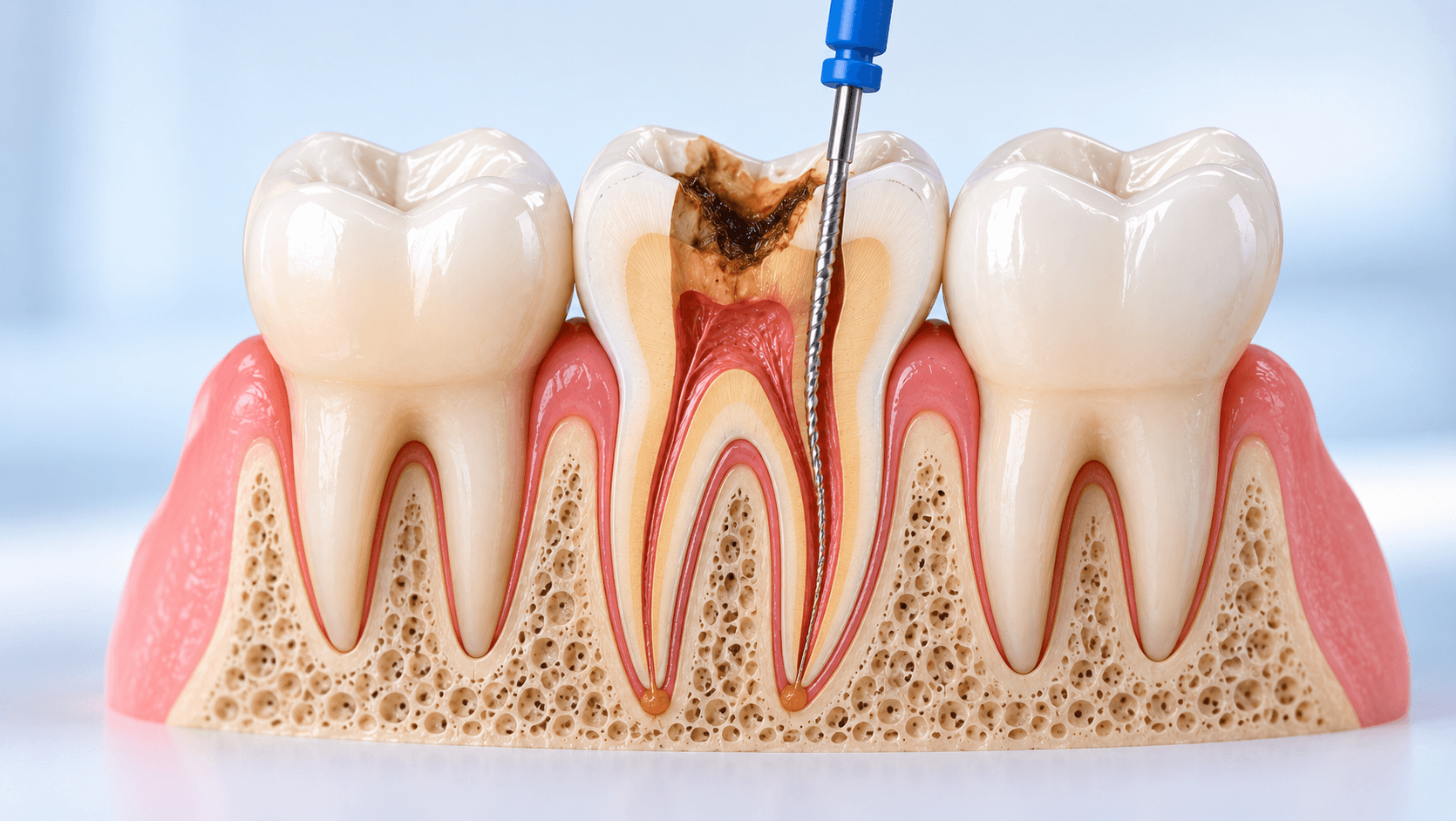
A patient returns eighteen months after a root canal with a sinus tract over the upper first molar. The radiograph shows a well-condensed, well-extended fill in three canals and a persistent periapical radiolucency. Nothing looks wrong, and yet something plainly is.
Endodontic failure is rarely mysterious. In the large majority of cases it reduces to one of a small number of causes, and most of them are anatomical or procedural rather than biological bad luck. This article covers what those causes are, how to identify which one you are looking at, and how to choose between retreatment, surgery and extraction.
Failure is a biological problem, not a radiographic one
Root canal treatment succeeds when the microbial load within the root canal system is reduced enough, and sealed well enough, that periapical inflammation resolves. It fails when viable bacteria persist in a space that communicates with the periradicular tissues, or when bacteria re-enter.
This framing matters because it explains why a radiographically beautiful root filling can fail and a radiographically unimpressive one can succeed. The radiograph shows the filled space. It does not show the unfilled space, and the unfilled space is where failure lives.
Every cause below is a variation on one of two themes: bacteria that were never removed, or bacteria that got back in.
Cause 1: Missed canals, and the MB2
The single most common anatomical cause of failure in the maxillary molar is an untreated second mesiobuccal canal.
The MB2 is present in a large majority of maxillary first molars - reported prevalence varies widely with method of investigation, but studies using magnification and troughing find it far more often than studies based on radiographs alone. It is also present frequently in maxillary second molars.
Other commonly missed anatomy includes the second distal canal in mandibular first molars, the middle mesial canal in mandibular molars, a second canal in mandibular incisors and premolars, and C-shaped configurations in mandibular second molars.
The practical conclusion: if you treat maxillary molars and you are not routinely finding an MB2, you are not routinely missing it because it is absent. Magnification, a modified access outline toward the mesiobuccal line angle, ultrasonic troughing of the developmental groove between MB1 and the palatal orifice, and patience are what change your detection rate. Our Advanced Endodontic Course covers MB2 location and molar access under magnification specifically because this is where most general practice endodontics is lost.
Cause 2: Inadequate disinfection
Instrumentation shapes; irrigation disinfects. A canal can be shaped to a large apical size and remain infected if the irrigation protocol is poor.
The common deficiencies are insufficient volume of sodium hypochlorite, inadequate contact time, a needle that cannot reach the apical third, no activation, and no attention to the isthmuses, fins and lateral anatomy that files never touch.
Mechanical instrumentation contacts only part of the canal wall in oval and irregular canals. Everything else depends on the irrigant reaching it, being replenished, and being active. Activation - ultrasonic, sonic or laser-assisted - and a sensible sequence including EDTA for the smear layer are not refinements; they are the part of the procedure that actually kills bacteria.
Intracanal medicament between visits has a role in cases with persistent exudate or symptoms, but it does not rescue a poor irrigation protocol.
Cause 3: Coronal leakage
This is the most avoidable failure of all. An endodontically excellent tooth left in a provisional restoration for months, or restored with a leaking composite, becomes recontaminated along the entire canal system.
The practical rules are simple and widely ignored. Place a definitive coronal seal promptly. Use rubber dam for the restorative phase as well as the endodontic phase. Protect the cusps of root-treated posterior teeth with cuspal coverage, because a tooth that fractures is lost regardless of the quality of the endodontics.
If you are deciding where to spend effort to improve your endodontic success rate, coronal seal gives a better return than almost anything else.
Cause 4: Procedural errors
Ledging, canal transportation and zipping move the prepared canal away from the original anatomy and leave infected dentine untouched.
Perforation - strip perforation in the furcal wall of curved roots, or crown perforation during access - creates a direct communication with the periodontium.
Separated instruments block access to the apical anatomy. The instrument itself is usually not the problem; what is left uncleaned beyond it is.
Over-instrumentation and extrusion of irrigant or filling material beyond the apex causes inflammation and in the case of sodium hypochlorite accidents, significant morbidity.
Under-extension leaves apical anatomy untreated. Over-extension does not compensate for it.
Cause 5: Vertical root fracture and cracks
Vertical root fracture carries the worst prognosis of all and is frequently misdiagnosed as a failed root filling.
The classic presentation is a narrow, deep, isolated periodontal pocket, sometimes with a sinus tract located more coronally than you would expect from an apical lesion, and a J-shaped or halo radiolucency along the root. CBCT may show it; often it does not, because the fracture is narrower than the voxel size.
Risk factors include excessive dentine removal during shaping, over-aggressive post space preparation, large posts, lateral condensation forces in thin roots, and unprotected cusps.
The reason this matters clinically is that retreatment of a vertically fractured root will fail, and the patient will have paid for it. If the signs point to a fracture, investigate properly - including a surgical flap to visualise the root if necessary - before committing the patient to retreatment.
Choosing between retreatment, surgery and extraction
Non-surgical retreatment is the default where the suspected cause is intracanal: a missed canal, inadequate disinfection, under-extension or coronal leakage. It addresses the actual source of infection. Access through an existing crown is usually preferable to removing a well-fitting restoration, provided the access does not compromise the restoration's retention.
Surgical endodontics is indicated where the canal system cannot be adequately re-accessed - a well-fitting post and core where removal risks fracture, a separated instrument that cannot be bypassed, a ledge that cannot be renegotiated - or where retreatment has already failed. Modern apical microsurgery with magnification, ultrasonic retropreparation and a biocompatible root-end filling material has considerably better outcomes than the older approaches it replaced.
Extraction becomes the rational choice with vertical root fracture, inadequate remaining tooth structure for a predictable restoration, insufficient ferrule, severe periodontal involvement, or a strategically unimportant tooth where implant or prosthetic alternatives are straightforward.
The decision should be driven by the identified cause of failure, not by a general preference for saving or replacing teeth.
Reducing your own failure rate
Three changes produce most of the improvement.
Magnification. The relationship between what you can see and what you can treat is direct. A microscope changes molar endodontics more than any file system.
Irrigation discipline. Volume, contact time, needle depth, activation, sequence. Write it down as a protocol and follow it.
Coronal seal and cuspal protection. Treat the restorative phase as part of the endodontic treatment, not as something that happens afterwards.
CBCT, used selectively, adds a great deal in failure assessment - identifying missed canals, confirming perforations, and assessing the extent of periapical lesions - without being necessary for routine primary treatment.
Our Advanced Endodontic Course covers molar RCT, MB2 location and retreatment hands-on, and our Online Advanced Endodontic Course covers the same material remotely. For case assessment, our Oral Radiology and CBCT courses address the interpretation side.
Related reading: our complete guide to rotary endodontics and our article on dental treatment planning.