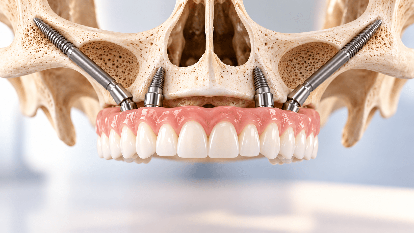
There is a category of patient every implant dentist eventually meets: the severely resorbed maxilla where conventional implants have nowhere to go, where sinus pneumatisation has consumed the posterior bone, and where the patient has already been told by two clinicians that they need eighteen months of grafting before anything can be done.
Zygomatic implants exist for that patient. They anchor in the zygomatic bone rather than the alveolus, bypassing the deficient maxilla entirely, and in suitable cases allow a fixed prosthesis to be loaded within days rather than after two years of staged reconstruction.
They are also among the least forgiving procedures in implant dentistry. This guide covers when they are indicated, how the protocols differ, what goes wrong, and what training is realistically required before attempting one.
When the maxilla is genuinely too atrophic
Classification systems help, but the clinical question is simpler: is there enough bone of adequate quality, in the right position, to place conventional implants that will survive loading?
In the posterior maxilla, pneumatisation of the sinus combined with vertical and horizontal resorption frequently leaves 1 to 3 mm of residual bone below the sinus floor. In the anterior maxilla, long-standing denture wear and buccal plate loss can leave a knife-edge ridge that will not accept a standard-diameter fixture without augmentation.
The conventional answer is reconstruction: sinus augmentation, onlay or block grafting, guided bone regeneration, sometimes iliac crest harvest. These work. But they add cost, morbidity, a donor site in some cases, and a treatment timeline measured in many months, during which the patient wears a tissue-borne denture over a healing graft.
Zygomatic implants offer a different trade: accept a technically demanding surgery once, and avoid the graft and the waiting entirely.
Anatomy that actually matters
The zygomatic bone is dense, consistently available, and remarkably stable in volume regardless of alveolar resorption. That is the whole premise.
The implant is long - commonly in the 30 to 55 mm range - and engages the zygomatic body. The critical anatomical structures along its path are the maxillary sinus and its membrane, the infraorbital nerve and foramen, the orbital floor, and the infratemporal fossa.
Two anatomical variables drive technique selection. First, the buccal concavity of the maxillary wall: a pronounced concavity means a classic intrasinus path would leave the implant body exposed within the sinus or perforating the wall. Second, the relationship of the zygomatic buttress to the planned prosthetic emergence, because an implant that is anatomically ideal but prosthetically palatal creates a bulky, unhygienic restoration the patient will hate.
The main surgical approaches
Classic intrasinus technique. The original approach, with the implant passing through the sinus from the alveolar crest to the zygoma. Simple in concept, but head position and the buccal concavity often force a palatal emergence.
Sinus slot technique. A window is prepared in the lateral sinus wall to guide the drill path while keeping the implant closer to the sinus wall, improving emergence.
Extramaxillary or extrasinus approach. The implant body runs outside the sinus along the lateral maxillary wall, with only the apex engaging the zygoma. This generally gives the best prosthetic emergence and avoids the sinus membrane, but leaves part of the implant body unsupported by bone and can produce a palpable or visible contour.
ZAGA, the zygoma anatomy-guided approach. Rather than committing to one technique, the path is selected according to the individual patient's maxillary wall anatomy, classified from a fully intrasinus to a fully extramaxillary configuration. This is now the mainstream way of thinking about the decision: the anatomy dictates the technique, not the operator's preference.
Quad zygoma, hybrids and pterygoid implants
Where the anterior maxilla also has insufficient bone for conventional implants, four zygomatic implants - two per side, commonly called quad zygoma - can support a full-arch fixed prosthesis without any conventional fixtures.
More often the arch is hybrid: two zygomatic implants posteriorly and two to four conventional implants anteriorly, where residual bone usually persists longest.
Pterygoid implants are a related graftless solution for the posterior maxilla, engaging the pterygoid plate. They are often discussed alongside zygomatic implants because they solve the same problem - posterior anchorage without sinus grafting - and because clinicians who adopt one usually end up learning both. Our zygomatic and basal implant courses cover both in a combined surgical sequence.
Loading protocol and prosthetic considerations
Immediate loading is common in zygomatic cases, and this is one of the attractions for patients. Primary stability in the dense zygomatic bone is usually high, and splinting multiple fixtures with a rigid provisional distributes load.
The prosthetic reality is more demanding than the surgical marketing suggests. Angulation is significant, so angled multi-unit abutments are routine. Emergence is often more palatal than ideal, which affects phonetics, tongue space and hygiene access. The prosthesis must be designed for cleansability from the outset, because a patient who cannot clean under a full-arch bridge supported by implants reaching into the zygoma will develop soft tissue problems that are difficult to resolve.
Plan the prosthesis first. Surgery that ignores the final restoration produces technically successful implants carrying a restoration nobody is happy with.
Complications you should expect to meet
Sinusitis is the most frequently reported late complication. It may arise from membrane perforation, from an oroantral communication along the implant path, or from compromised sinus drainage. Pre-existing sinus pathology should be assessed and treated before surgery, and patients with chronic sinus disease need careful counselling.
Soft tissue recession and peri-implant mucositis around the zygomatic emergence, often related to thin keratinised tissue and difficult hygiene access.
Oroantral communication, requiring closure.
Orbital penetration - rare, catastrophic, and entirely avoidable with proper planning and drilling control.
Infraorbital nerve injury, with paraesthesia of the cheek and upper lip.
Implant failure, which in this context is a far more serious problem than losing a conventional implant, because the salvage options are limited and the patient has usually already exhausted conventional alternatives.
Planning with CBCT is not optional. Neither is a frank discussion with the patient about what failure would mean.
What training is actually required
Zygomatic implant surgery is not a procedure to add after a weekend course, and it is not a natural next step immediately after basic implantology. A realistic progression looks like this.
First, competence in conventional implant placement including the posterior maxilla, with enough case volume to have met and managed complications.
Second, competence in sinus procedures - lateral window augmentation and the management of membrane perforation - because you must understand the sinus before you traverse it.
Third, full-arch rehabilitation experience, including immediate loading protocols and full-arch prosthetic design.
Fourth, formal zygomatic training with cadaver or model work, CBCT planning practice, and ideally supervised or mentored first cases.
Our Zygomatic Implant Course in India covers hands-on zygomatic implantology, and our Advanced Dental Implant Course covers the bone grafting, sinus lift and guided surgery foundations that should precede it. For the prosthetic side, our Implant Prosthodontics Course addresses angled multi-unit workflows and full-arch design.
Related reading on this site: our comparison of basal implants and conventional dental implants, and our complete guide to modern implant dentistry.